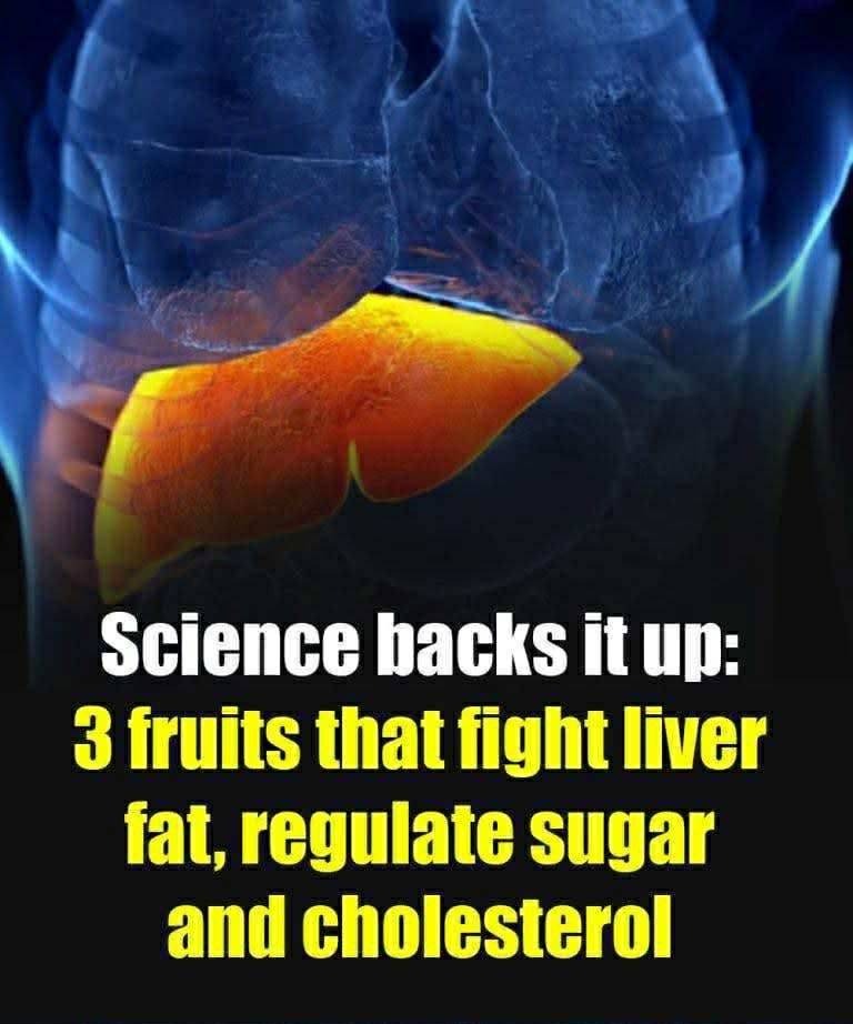
From Pathophysiology to Phytochemicals: Leveraging Specific Fruits for Liver Health
Non-Alcoholic Fatty Liver Disease (NAFLD), recently rebranded as Metabolic Dysfunction-Associated Steatotic Liver Disease (MASLD), represents a global health crisis. Characterized by the excessive accumulation of triglycerides (fat) within liver cells (hepatocytes), MASLD is the hepatic manifestation of the modern metabolic syndrome. It is intimately and causally linked to insulin resistance, central obesity (visceral fat), Type 2 Diabetes, hypertension, and dyslipidemia (abnormal cholesterol).
ad
The hopeful reality is that, unlike many chronic conditions, MASLD is often highly responsive—and potentially reversible—through targeted lifestyle and dietary interventions. Clinical consensus and robust research now confirm that strategic dietary changes can induce significant liver fat reduction, often quantified at a 10–15% reduction in liver fat within a 12-week period, which correlates with major improvements in metabolic function.
This extensive analysis will dissect the pathophysiology of MASLD and provide an exhaustive scientific exploration of the specific mechanisms—the phytochemicals, fibers, and fatty acid profiles—of blueberries, apples, and avocados, demonstrating how these three fruits serve as cornerstones in a liver-healing diet.
Part I: 🔬 The Pathophysiology of MASLD (NAFLD)
Understanding the mechanisms driving MASLD is crucial for appreciating how diet works to reverse it. The disease progresses through a series of metabolic failures.
1. The Genesis: Insulin Resistance and De Novo Lipogenesis (DNL)
MASLD begins not in the liver, but in the fat and muscle tissues, which become resistant to the action of insulin.
Insulin’s Failure: Insulin is the key hormone that instructs cells to take up glucose. When cells become resistant, the pancreas compensates by secreting excessive insulin (hyperinsulinemia).
Adipose Overflow: Insulin resistance in adipose (fat) tissue leads to uncontrolled breakdown of stored fat (lipolysis), flooding the bloodstream with free fatty acids (FFAs).
The Liver’s Burden: These excess FFAs travel to the liver. Simultaneously, the hyperinsulinemia stimulates the liver enzyme pathways responsible for De Novo Lipogenesis (DNL)—the process of synthesizing fat directly from excess carbohydrates (glucose and fructose). The liver is forced to take in and create fat simultaneously, overwhelming its capacity and leading to hepatic steatosis (fatty liver).
2. The Progression: Oxidative Stress and Inflammation (The “Second Hit”)
Steatosis, while reversible, can progress to the more dangerous stage of Nonalcoholic Steatohepatitis (NASH)—a condition marked by fat accumulation plus inflammation and cell death.
Mitochondrial Dysfunction: The excessive fat stored in the liver forces the mitochondria (the cell’s powerhouses) to work overtime to try and burn it. This metabolic overload causes mitochondrial dysfunction, leading to the production of Reactive Oxygen Species (ROS), or free radicals.
Oxidative Stress: This ROS production is termed oxidative stress. It damages hepatocyte membranes, DNA, and proteins.
Cytokine Cascade: The damaged and dying cells trigger the release of inflammatory molecules (cytokines), particularly from resident immune cells (Kupffer cells). This systemic inflammation perpetuates the cycle, eventually leading to fibrosis (scarring), and potentially cirrhosis (advanced, irreversible scarring).
3. Key Risk Factors and Co-Morbidities
The risk of developing MASLD is exceptionally high in individuals presenting with key metabolic imbalances:
Visceral Adiposity: Carrying excess weight specifically around the stomach (visceral fat) is a primary indicator. Visceral fat is hormonally active and highly inflammatory.
Type 2 Diabetes and PCOS: Both conditions are characterized by chronic, systemic insulin resistance.
Dyslipidemia and Hypertension: High triglycerides, low HDL, and high blood pressure reflect the underlying vascular and metabolic dysfunction driven by the fat accumulation.
Part II: 🫐 Blueberries – Anthocyanins and Metabolic Control
Blueberries (and other dark berries) are therapeutic agents in MASLD due to their incredibly high concentration of anthocyanins and their role in modulating core fat and glucose pathways.
1. The Chemical Power of Anthocyanins
Anthocyanins are polyphenolic pigments responsible for the deep blue, purple, and red colors found in berries. They are potent signaling molecules in the body.
Direct Antioxidant Action: Anthocyanins stabilize cell membranes against ROS damage, directly reducing the oxidative stress that characterizes the inflammatory progression of MASLD. They neutralize free radicals before they can attack the hepatocyte.
Modulation of Fat Metabolism (Lipid Oxidation): Studies utilizing imaging (MRI or ultrasound) show that daily blueberry consumption correlates with reduced liver fat. Mechanistically, anthocyanins work by:
Inhibiting Lipogenesis: Downregulating key enzymes (like Fatty Acid Synthase) involved in synthesizing fat from carbohydrates.
Promoting Beta-Oxidation: Upregulating genes and enzymes (like AMPK) that encourage the liver cell to burn (oxidize) existing stored fat for energy.
Insulin Sensitivity and Glucose Homeostasis: Anthocyanins enhance insulin signaling pathways, resulting in better peripheral glucose uptake. This increased insulin sensitivity reduces the need for the pancreas to secrete high levels of insulin (hyperinsulinemia), thereby reducing the primary driving force of hepatic DNL.
2. Gut Health and Systemic Inflammation
Blueberries’ benefits extend beyond direct action on the liver; they promote a healthier metabolic environment via the gut-liver axis.
